As many of you know last week a team of the University of Leicester have publicly revealed to have discovered, in all likelihood, the tomb of Richard III. The results seem comforted by the analysis of mitochondrial DNA, while the discrepancy on the Y chromosome could be explained by a false paternity. The study was completed with a forensic facial reconstruction of the king, performed by the experts of the University of Dundee, led by Caroline Wilkinson, Professor of Craniofacial Identification.
Given the opportunity, I decided to publish here our state of the art on this particular field (forensic facial reconstruction applied to archeology), publishing the presentation that I gave during the study day in honor of Prof. Franco Ugo Rollo (Ascoli Piceno, November 26 2014).
... and here is a brief explanation of each slide:
SLIDE 1
A remember of Franco Ugo Rollo, professor at the Camerino University. It was not my fortune to know personally Prof. Rollo, but his name is surely well known also in my discipline (archeology).
SLIDE 2
"Digital faces: new technologies for the forensic facial reconstruction of the historical figures".
The presentation intend to be an overview of the digital methodologies of FFR with FLOSS, developed in the last two years on the blog ATOR with a spontaneous contribution of different authors.
SLIDE 3
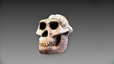
The traditional work-flow involves several operations: 3D scanning the skull, preparing a replica, performing the anthropological analyses, placing the tissue depth markers, reconstructing the profile, modeling the muscles and skin, calibrating the model with the available sources and dressing it.
SLIDE 4
The same operations are necessary for the digital work-flow. Our main work has been to turn the traditional process into a digital one, using only FLOSS.
SLIDE 5
There are different technology to obtain a 3D digital copy of the original skull. The main two we are using are: SfM - IBM and X-ray CT.
SLIDE 6
IN 2009 Arc-Team perform the first test in applying SfM - IBM with FLOSS to Cultural Heritage, during its participation at the TOPOI excelent cluster of Berlin.
SLIDE 7
The test developed in a collaboration with the French researcher +Pierre Moulon (Université Paris - Est and Mikros Image; actually at Acute3D) to integrate SfM - IBM software in ArcheOS 4 (codename Caersar)
SLIDE 8
The first test (TOPOI Löwe) gave positive results
SLIDE 9
The process is mainly based on different photos with different orientations, computing the displacement of common points between images
SLIDE 10
To complete the 3D documentation of an object, the next step is the so-called mesh-editing, which can be performed in the software MeshLab (developed by the Visual Computing Lab at the ISTI - CNR of Pisa, Italy)
SLIDE 11
In order to validate the digital method of FFR, some unconventional procedures (derived from the hacker culture) have been adopted. With reverse engineering techniques, based on SfM, it has been possible to digitally replicate the process of past FFR projects and to compare the results.
SLIDE 12
The anthropological validation has been performed comparing the result of 3D models obtained with SfM - IBM and the relative results coming form 3D scan (the observed distortion remained in the range of 1 mm).
SLIDE 13
In several projects it is possible to work with DICOM data. In these cases the anthropological analysis is more accurate. (3D VS Voxel)
SLIDE 14
The main software we used for DICOM data is InVesalius, mainly developed at the Renato Archer Information of Technology Center, an institute of the Brazilian Ministry of Science and Technology.
SLIDE 15
"X-ray computed tomography (X-ray CT) is a technology that uses computer-processed X-rays to produce tomographic images (virtual 'slices') of specific areas of the scanned object, allowing the user to see inside without cutting." (Wikipedia)
SLIDE 16
Also in this case, the process was validated with unconventional procedures derived from hacker culture. With reverse engineering of CT videos it has been possible to rebuild DICOM data and the 3D model of different skulls, replicating FFR projects and comparing the results.
SLIDE 17
It is necessary to check and validate the protocol with a continuous methodological comparisonwith all the available resources. For this reason, we tried also the FFR of Henry the IV, a project in which Prof. Rollo was involved, rejecting (with other scholars) the attribution of the mummified head to the French king. Our test in this case is just an experiment, starting from low quality data, but it is a good example to show some benefits of digital FFR, like the possibility to quickly modify the reconstructed face (e.g. closing the mouth in order to perform superimposition with the death mask), an operation not so simple with tangible models.
SLIDE 18
Once obtained the 3D model, digital anthropological analyses do not differ from traditional ones.
SLIDE 19
In some cases, a virtual restoration of the model is necessary. The solution comes from symmetrical and boolean operations of 3D modeling software (Blender).
SLIDE 20
The whole process of 3D modeling is actually performed in the software Blender.
SLIDE 21
The first operation is to fix the 3D skull on the Frankfurt plane, which replicates the head position of a standing human figure.
SLIDE 22
Than tissue depth markers are placed. The software keeps automatically the correct normal of each marker.
SLIDE 23
In our works, for depth tissue markers, we use the tables of Degreef et alii (2006)
SLIDE 24
A second step is the profile reconstruction.
SLIDE 25
For nose shape we refer to G. Lebedinskaya method.
SLIDE 26
The validation of the method came mainly from the comparison between FFR models and the facial DICOM data of living people, a simple simple with digital techniques, using the software CloudCompaer. All this experiment were conducted ans blind test (the artist did not know the identity and the fisionomy of the people).
SLIDE 27
According to the blind test, main deviations were detected on the cheeks.
SLIDE 28
Like other 3D operations, muscles modeling has been performed in Blender.
SLIDE 29
The technique hes been continuously rationalized and optimize. For instance, once the main muscles are modeled with metaballs in Blender, the result can be reused in successive reconstructions through an anatomical deformation.
SLIDE 30
It is possible to reach more realistic results through specific modeling tools,
like the "sculpt mode" in Blender.
SLIDE 31
Also skin modeling is an operation to be performed in Blender
SLIDE 32
Again the technique has been optimized: In order to simplify and speed up the process, a neutral facial model has been created.
SLIDE 33
The neutral model can be anatomically deformed on different skulls to meet gender and age dimorphism.
SLIDE 34
At the same time, the neutral model can be deformed to meet the anatomical criteria which determine the individual dimorphism.
SLIDE 35
After the reconstruction process, two main models are defined: one with hair and one hairless.
SLIDE 36
Thanks to the latest developments of the software MakeHuman it is now possible to further simplify and speed up the technique. Our actual research is following this direction.
SLIDE 37
The first tests carried out in 2014 have yielded positive results, thanks to the new feature which loads base raster images. The software is also perfectly compatible with Blender.
SLIDE 38
A further development of the protocol will allow to obtain high quality forensic facial reconstructions, in less time, without the need to master the techniques of 3D modeling.
SLIDE 39
At the end of the FFR process, the final model is calibrated with historical, archaeological and medical sources.
SLIDE 40
In case of historical reconstructions, the model appearance (hairstyle and clothing) is calibrated depending on era and culture, while the physical characteristics (color of hair and eyes) are set basing on the ancestry.
SLIDE 41
The 3D printing technologies allow the materialization of the model with different levels of detail.
SLIDE 42
A case study: the forensic facial reconstruction of St. Anthony of Padua
SLIDE 43
The 3D scan was carried out on the bronze cast performed by R. Cremesini in 1981.
SLIDE 44
The cast done by R. Cremesini is very important, because it derives from the temporary anatomical reconnection of the skull and the jaw, which were separated since the first survey of the tomb (1263).
SLIDE 45
3D scan has been performed with the SfM - IBM software of the archaeological GNU/Linux distribution ArcheOS.
SLIDE 46
The final model has been presented Tuesday, June 10 at the event "Scoprendo il volto di Antonio" at the Centro Culturale S. Gaetano in PAdua (Italy)
SLIDE 47 - 50
Digital FFR allows to further define the details of the model to reach a more realistic result.
SLIDE 51
Thanks to the collaboration with the Centro de Tecnologia da Informação Renato Archer - CTI (Ministério da Ciência and Technology do Brasil) the model was printed in 3D.
SLIDE 52
One of the materialized models was repainted by the Brazilian Mari Bueno,
specialized in religious art.
SLIDE 53
Thank you for your attention!
... and here is a brief explanation of each slide:
SLIDE 1
A remember of Franco Ugo Rollo, professor at the Camerino University. It was not my fortune to know personally Prof. Rollo, but his name is surely well known also in my discipline (archeology).
SLIDE 2
"Digital faces: new technologies for the forensic facial reconstruction of the historical figures".
The presentation intend to be an overview of the digital methodologies of FFR with FLOSS, developed in the last two years on the blog ATOR with a spontaneous contribution of different authors.
SLIDE 3
The traditional work-flow involves several operations: 3D scanning the skull, preparing a replica, performing the anthropological analyses, placing the tissue depth markers, reconstructing the profile, modeling the muscles and skin, calibrating the model with the available sources and dressing it.
SLIDE 4
The same operations are necessary for the digital work-flow. Our main work has been to turn the traditional process into a digital one, using only FLOSS.
SLIDE 5
There are different technology to obtain a 3D digital copy of the original skull. The main two we are using are: SfM - IBM and X-ray CT.
SLIDE 6
IN 2009 Arc-Team perform the first test in applying SfM - IBM with FLOSS to Cultural Heritage, during its participation at the TOPOI excelent cluster of Berlin.
SLIDE 7
The test developed in a collaboration with the French researcher +Pierre Moulon (Université Paris - Est and Mikros Image; actually at Acute3D) to integrate SfM - IBM software in ArcheOS 4 (codename Caersar)
SLIDE 8
The first test (TOPOI Löwe) gave positive results
SLIDE 9
The process is mainly based on different photos with different orientations, computing the displacement of common points between images
SLIDE 10
To complete the 3D documentation of an object, the next step is the so-called mesh-editing, which can be performed in the software MeshLab (developed by the Visual Computing Lab at the ISTI - CNR of Pisa, Italy)
SLIDE 11
In order to validate the digital method of FFR, some unconventional procedures (derived from the hacker culture) have been adopted. With reverse engineering techniques, based on SfM, it has been possible to digitally replicate the process of past FFR projects and to compare the results.
SLIDE 12
The anthropological validation has been performed comparing the result of 3D models obtained with SfM - IBM and the relative results coming form 3D scan (the observed distortion remained in the range of 1 mm).
SLIDE 13
In several projects it is possible to work with DICOM data. In these cases the anthropological analysis is more accurate. (3D VS Voxel)
SLIDE 14
The main software we used for DICOM data is InVesalius, mainly developed at the Renato Archer Information of Technology Center, an institute of the Brazilian Ministry of Science and Technology.
SLIDE 15
"X-ray computed tomography (X-ray CT) is a technology that uses computer-processed X-rays to produce tomographic images (virtual 'slices') of specific areas of the scanned object, allowing the user to see inside without cutting." (Wikipedia)
SLIDE 16
Also in this case, the process was validated with unconventional procedures derived from hacker culture. With reverse engineering of CT videos it has been possible to rebuild DICOM data and the 3D model of different skulls, replicating FFR projects and comparing the results.
SLIDE 17
It is necessary to check and validate the protocol with a continuous methodological comparisonwith all the available resources. For this reason, we tried also the FFR of Henry the IV, a project in which Prof. Rollo was involved, rejecting (with other scholars) the attribution of the mummified head to the French king. Our test in this case is just an experiment, starting from low quality data, but it is a good example to show some benefits of digital FFR, like the possibility to quickly modify the reconstructed face (e.g. closing the mouth in order to perform superimposition with the death mask), an operation not so simple with tangible models.
SLIDE 18
Once obtained the 3D model, digital anthropological analyses do not differ from traditional ones.
SLIDE 19
In some cases, a virtual restoration of the model is necessary. The solution comes from symmetrical and boolean operations of 3D modeling software (Blender).
SLIDE 20
The whole process of 3D modeling is actually performed in the software Blender.
SLIDE 21
The first operation is to fix the 3D skull on the Frankfurt plane, which replicates the head position of a standing human figure.
SLIDE 22
Than tissue depth markers are placed. The software keeps automatically the correct normal of each marker.
SLIDE 23
In our works, for depth tissue markers, we use the tables of Degreef et alii (2006)
SLIDE 24
A second step is the profile reconstruction.
SLIDE 25
For nose shape we refer to G. Lebedinskaya method.
SLIDE 26
The validation of the method came mainly from the comparison between FFR models and the facial DICOM data of living people, a simple simple with digital techniques, using the software CloudCompaer. All this experiment were conducted ans blind test (the artist did not know the identity and the fisionomy of the people).
SLIDE 27
According to the blind test, main deviations were detected on the cheeks.
SLIDE 28
Like other 3D operations, muscles modeling has been performed in Blender.
SLIDE 29
The technique hes been continuously rationalized and optimize. For instance, once the main muscles are modeled with metaballs in Blender, the result can be reused in successive reconstructions through an anatomical deformation.
SLIDE 30
It is possible to reach more realistic results through specific modeling tools,
like the "sculpt mode" in Blender.
SLIDE 31
Also skin modeling is an operation to be performed in Blender
SLIDE 32
Again the technique has been optimized: In order to simplify and speed up the process, a neutral facial model has been created.
SLIDE 33
The neutral model can be anatomically deformed on different skulls to meet gender and age dimorphism.
SLIDE 34
At the same time, the neutral model can be deformed to meet the anatomical criteria which determine the individual dimorphism.
SLIDE 35
After the reconstruction process, two main models are defined: one with hair and one hairless.
SLIDE 36
Thanks to the latest developments of the software MakeHuman it is now possible to further simplify and speed up the technique. Our actual research is following this direction.
SLIDE 37
The first tests carried out in 2014 have yielded positive results, thanks to the new feature which loads base raster images. The software is also perfectly compatible with Blender.
SLIDE 38
A further development of the protocol will allow to obtain high quality forensic facial reconstructions, in less time, without the need to master the techniques of 3D modeling.
SLIDE 39
At the end of the FFR process, the final model is calibrated with historical, archaeological and medical sources.
SLIDE 40
In case of historical reconstructions, the model appearance (hairstyle and clothing) is calibrated depending on era and culture, while the physical characteristics (color of hair and eyes) are set basing on the ancestry.
SLIDE 41
The 3D printing technologies allow the materialization of the model with different levels of detail.
SLIDE 42
A case study: the forensic facial reconstruction of St. Anthony of Padua
SLIDE 43
The 3D scan was carried out on the bronze cast performed by R. Cremesini in 1981.
SLIDE 44
The cast done by R. Cremesini is very important, because it derives from the temporary anatomical reconnection of the skull and the jaw, which were separated since the first survey of the tomb (1263).
SLIDE 45
3D scan has been performed with the SfM - IBM software of the archaeological GNU/Linux distribution ArcheOS.
SLIDE 46
The final model has been presented Tuesday, June 10 at the event "Scoprendo il volto di Antonio" at the Centro Culturale S. Gaetano in PAdua (Italy)
SLIDE 47 - 50
Digital FFR allows to further define the details of the model to reach a more realistic result.
SLIDE 51
Thanks to the collaboration with the Centro de Tecnologia da Informação Renato Archer - CTI (Ministério da Ciência and Technology do Brasil) the model was printed in 3D.
SLIDE 52
One of the materialized models was repainted by the Brazilian Mari Bueno,
specialized in religious art.
SLIDE 53
Thank you for your attention!