Wednesday, 29 July 2015

When Veterinary Medicine and 3D printing meet each other


TV story with English subtitle


The Brazilian Team of Forensic Anthropology and Forensic Dentistry (Ebrafol) was founded in 2014. Comprising a number of liberal professionals, mostly belonging to the field of Dentistry, it always sought a shortcut using the know-how of its members and considering the necessity of the Brazilian population for relevant applications. Even before the Ebrafol started, we (me and Dr. Paulo Miamoto) had already worked a handful of partnerships and they would contemplate not only the human population but also other animals.

TV Story with English subtitle
In the second half of 2013 we have met the Veterinarian Dr. Roberto Fecchio. He was well known for his mastery in saving the lives of many animals and bringing dignity and quality of life to them. Rebuilt beaks, perfectly fitting prosthesis, and implanted well treated teeth. And I mean animals ranging from a small rodent to a scary feline, whether a Guinea pig or a lion, there would be Dr. Fecchio and his staff, caring for and rehabilitating them.
When I meet Dr. Fecchio (at center) at Sao Paulo University (USP)

It seems a short period, but since 2013 a lot has happened. In the meantime, regarding skills in computer graphics applied to human and animal health, our knowledge advanced quite a bit. Since 2013, Dr. Fecchio would motivate us to develop 3D-printed prosthetic beaks, but at that time we just did not have the necessary know-how to actually model them, nor the equipment to print them.

The red-footed tortoise (Chelonoidis carbonaria) "Fred" in the surgery home

That changed a few weeks ago. Dr. Paulo Miamoto purchased a 3D printer. The goal was to explore it for scientific studies and commercial printing. All was very new, interesting and unknown.

Wealthy tortoise scanned (wireframe) and Fred inside it.

Upon learning about the 3D printer, Dr. Fecchio, always at the forefront, proposed that we participated in a project with him, from Santos-SP, and other team, from Brasília-DF. The case was about a poor tortoise, who had been the victim of a bushfire in the Brazilian plains. The flames injured her hoof and she lost a considerable part of its structure. Luckily the animal was rescued and taken barely alive to the hands of Dr. Rodrigo Rabello, whom with the aid of his brother Dr. Matheus Rabello, successfully treated and healed two pneumonia episodes and other diseases caused by the animal’s deficient immune system.

System of matching

Although the tortoise regained stable health, she tortoise found herself in big trouble. She had no hoof as the bony plates that were left fell off and gave her a shelled egg-like aspect, with only a thin membrane which could be perforated quite easily.

Hoof exploded

That’s when Dr. Fecchio stepped in, proposing the partnership and finding himself quite content with everybody’s agreement to participate in this project.

I figured the reconstruction of the hoof could be made using a simple methodology. First we would do a 3D scan of the tortoise who had lost the hoof. The technique used is called photogrammetry. Roughly, we took several pictures of the animal, sent them to a computational algorithm and it reconstructed the 3D volume. Then we did the same with a healthy tortoise hoof. This way, we digitized the 3D volumes of the tortoise without a hoof and the healthy hoof. Then, we would just have to proceed with Boolean calculations and, a structure that fits the sick animal is obtained.

Printed part

Of course we had a lot of problems in the process. The hoof had to be printed in 4 parts because we did not know if Dr. Paulo’s printer would finish the job in time. That’s why we divided it into four pieces, so that we could hire companies or people who offered this service in case we had any problems with 3D printing. Fortunately, 3D prints were successful, although this process wasn’t quite quick. It took five days of almost uninterrupted printing for the hoof to get ready. After that, we had an unpleasant surprise upon cleaning the support material created by the printer. In the joint areas it was very difficult to remove it. Thanks to the help of Dr. Paulo Esteves, an experienced Dentist, cleaning the support material was possible and everything went smoothly well.

Te team after surgery (I'm on the grayscale photo)

The surgery was covered by the largest Brazilian TV station, Rede Globo. The procedure was a success and at the end Fred the tortoise, received a new hoof and it wasn’t necessary to screw it to bony parts of her body, as a photogrammetry provided a high precision scanning of the area and made possible a very nice adaptation of the prosthesis.

Steps of surgery - toucan

Meanwhile, another case had been handled by the team. Zeca, The toucan, broke his beak when he hit a window. A homologous prosthesis was installed using a cadaver beak adapted to the fracture, which is a common practice in veterinary medicine. Unfortunately, Zeca’s “new” beak could not stand a very high load and broke. Upon seeing that the toucan had lost his beak, Dr. Fecchio proposed reviving the first project that we developed together, back in the pre-Ebrafol period, i.e., to create digitized beak prosthesis. Inspired by the successful surgery, we got back in track and to our complete joy, everything worked out and Zeca is fully adapted to his new beak!

Our team is very happy and honored for all that has happened. Besides the feeling of nobility and accomplishment, we are also proud of accomplishing everything using free and open software. Photogrammetry was done with PPT-GUI, and 3D modeling was done in Blender. We used Cork for Boolean calculations and sliced the mesh for printing with Slic3r.

We barely enjoyed the taste of success and we are already engaged in a new project. Soon we'll post more news, see you!

Acknowledgements

Dr. Everton da Rosa, which made possible my trip to Brasilia to meet the Rabello doctors and participate in Fantástico, Brazil’s most popular Sunday TV show. Claudio Marques Sampaio (patola) to help us with 3D printing. Denise Oltramari, which provided us with one of the tortoises she takes care of for photogrammetry. Dr. Gilbert Bernstein to develope the powefull Cork, the boolean standalone used to create the prosthesis. Daniel Ludwig and Lis Caroline for the aid in the process of photography (photogrammetry). Giovanna Leite Soares and Dr. Paulo Miamoto, who assisted us with translations into English. To all the news crews that documented this project while respecting the scientific aspects and highlighting the importance of such initiatives for the sake of animals.
Tuesday, 28 July 2015

The meaning of an "open source exhibition"

As many of you know, since more than one year we are working on the exhibition "Facce. I molti volti della storia umana". Now that the exhibition was inaugurated and our work is completed, it is time to share with open licenses (CC-BY) what we produced. 
It will be a long process, as the materials are different (images, photos, video, 3D models), nevertheless we have to start uploading the documents. Thanks to a short discussion with +Maurizio Napolitano (Fondazione Bruno Kessler) and +Rodrigo Padula (Grupo Wikimedia Brasileiro de Educação e Pesquisa), both expert in open data, I think that the best solution will be to upload the data directly in ATOR, where I can cite all the people who participated in the "production process", from 3D scan to facial reconstruction, till scientific validation.
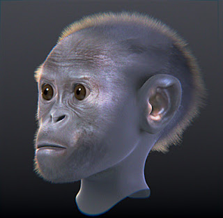
IMHO the best image to start with is the one of the Taung Child, for different reason: it has been the first attempt performed by our team in order to reconstruct the face of an hominid; it summarizes our concept of Open Research; it has been one of the ideas that gave birth to exhibition "Facce", as Dr. Nicola Carrara conceived it; it is the first project in which Arc-Team, the Anthropological Museum of the University of Padua and Antrocom worked together; last but not least, it is a perfect example of what we mean of open data. Indeed the first reconstruction we produced (version 1.0), which actually is already part of the related article in Wikipedia, has been modified after the development (and the validation) of a new technique of paleo-art, based on the anatomical deformation of a CT scan of a Pan troglodytes. For this reason now we have a new and more accurate reconstruction, which can be considered a version 2.0 of the same model. 
The open data we intend to share here in ATOR are meant to be open not only in the direction of free access for everyone, but also (most important) under the temporal dimension: they should just represent a step of a continuous evolution of the research, in which all the reconstructions can be considerate simply as the latest release of a model (exactly like in software development, with new versions and forks). For this reason we choose the Creative Commons Attribution license, in order to allow derived works and projects.
The two images below can explain better this concept: the first one represent the Taung Child reconstruction in his first version (based on an anatomical study of primates), while the second one is derived by the anatomical deformation of Pan troglodytes CT scan.

The first version of the reconstruction of the Taung Child

The second version of the Taung Child
Both of the models are the result of a team work, although most of the process (and in particular the most important and delicate phases) has been performed by the 3D artist  +Cícero Moraes. Here below I want to cite the credits for this reconstruction (following the same order of the work-flow):

1. 3D scan of the cast: Luca Bezzi (Arc-Team) and +Moreno Tiziani (Antrocom)
2. 3D modeling (skull restoration, anatomical study, CT deformation): +Cícero Moraes (Arc-Team)
3. scientific validation: Prof. Telmo Pievani (University of Padua, Department of Biology) and Dott. Nicola Carrara(Anthropological Museum of the University of Padua)

Acknowledgement:

The Anthropological Museum of the University of Pauda, for providing the cast of the fossil.
The KUPRI, Primate Research Institute Kyoto University, for sharing the CT scans of different primates.
Dr. Claudio Paluani (University of Padua), who, during the lesson "Digital bones" at the Botanical Garden of the University of Padua, had the same idea we had about the validation of the methodology of anatomical deformation through the modification of two CT scans of living primates. This fact convinced us to perform the test, after seeing that more people reached the same conclusion about the validation problem.

Monday, 27 July 2015

Documentation of a bas-relief on a cliff : the workflow

This summer, between May and June, we worked for a joint mission, led by the University of Innsbruck (Institut für Alte Geschichte und Altorientalistik) and the Cultural Heritage, Handcrafts and Tourism Organization of Iran. The project was held in Firuzabad, in the Pars Province of Iran. We will write more details about this work in the next post. By now I just want to use some material we collected to illustrate the work-flow in data acquiring during an archaeological documentation of a bas-relief on a cliff.
The video below shows the overall process.


You can see the initial preparation phase (1), during which we placed the Ground Control Point (GCP) to perform normal 2D vertical photo-mapping and to rectify and georeference the 3D point-cloud. Than (2) we collected pictures with three different flights of our DIY drone, in order to use them with different open source SfM/MVSR software (PPT, openMVG and MicMac), to reach the best possible result: a couple of flights with parallel camera, to have a good superimposition of the whole bas-relief, and a higher acquisition to cover the upper details. In the meantime (3) another operator (+Rupert Gietl) was collecting pictures from the ground, to register also the lower perspective. Later (4), I prepared the total station and collected the GCP, thanks to some fixed points we placed the day befor (0) with our GPS. Finally +Rupert Gietl  took the last (very close) details photos, using a ladder.
The entire process lasted more or less four hours, but we needed some more time the day before to place the fixed GCP down in the valley (in international Geographic Coordinates System). A good part of the work involved just the logistics or the approach to the site, and has been slowed by the transportation of the necessary equipment (ladder, total station and drone) through a couple of passages where it was necessary to climb some rocks.
It is interesting to note that it would not have been possible to accomplish this mission with a commercial drone, due to the embargo rules (which are currently under revision), while with a DIY hexacopter it has been simple to disassemble the components which were not allowed (like the FPV system ore the GPS controlled flight).
I hope this post was useful, have a nice day!

Thursday, 2 July 2015

The archaeometric excavation

Last year, on November 28, Arc-Team joined the conference "Lo scavo archeometrcio: scienza e tecnologia applicate allo scavo archeologico" (en: "The archaeometrcic excavation: science and technology applied to the archaeological excavation"), which was held in Rovereto (Italy) at the Museo Civico.
During the meeting we gave a presentation titled "Professional archaeology. Innovations and best practice with free technology. Toward an Open Research." Today I uploaded on our server the slides, so that we can share this work (like always under Creative Commons Attribution - CC BY).
As usual the presentation has been done with impress.js through the Graphical User Interface Strut (both GPL licensed) and it is optimized for Firefox or Iceweasel (better visualized here).


Here is a little explanation regarding the single slides:

SLIDE 1
A fast presentation regarding Arc-Team.
SLIDE 2
An animation representing the importance of geocoding in archaeology (from space to site).

SLIDE 3
Differential GPS and Total Station: the main tools needed by archaeologists on the field (to georeference every single element of the archaeological record).

SLIDE 4
Some examples of geocoding in archaeology: everyday work, project in extreme conditions and missions abroad...

SLIDE 5
... survay and excavations

SLIDE 6
In survay projects the geocoding tolerance for archaeology is higher, so that we are testing alternative solutions to build a low-cost and open source GPS with centimetric accuracy, using the software RTKLIB (or its port in Android)

SLIDE 7
All the recorded data (in 2D and 3D) can be imported into an open source GIS.

SLIDE 8
For aerial archaeology it since 2008 we are working with open source DIY UAV, like the UAVP or the KKcopter (in the slide).

SLIDE 9
Our last UAV prototype and an example of 3D pointcloud form aerial pictures.

SLIDE 10
Since 2014 we are testing DIY camera (using the filter of Public Lab) for NDVI and NGB pictures in archaeological remote sensing.

SLIDE 11
Just removing the IR filter, a normal camera can be used for endoscopic prospections in low light conditions.

SLIDE 12
In the field of geophysical prospections we use a DIY  machine for Electrical Resistivity Imaging. The data can be visualized in a GIS (e.g. GRASS GIS in the slide), using the east and north and the resistivity values.

SLIDE 13
Some geoarchaeological analyses can be performed directly on the field, like the settlement test (using the soil triangle) for the texture or the lithologic recognition for the skeleton.

SLIDE 14
Also some basic analytical chemistry can help during the excavation (giving indications on the ancient use of the soil), to verify the presence/absence of phosphates or of organic remains.

SLIDE 15
Other preliminary laboratory (flotation and sieving) analyses can prepare the samples for further investigation. Also in this case we use a DIY machine.

SLIDE 16
Colorimetry can be performed in many ways. Currently we are testing different options, like the open source spectrometer of Public Lab.

SLIDE 17
For some laboratory geoarchaeological analysis (e.g. microscopic morphology) we use normal optic microscopes, while for more advanced studies we externalize the service (e.g. SEM or energy dispersive x-ray spectroscopy)

SLIDE 18
Currently we are testing the potentialities of the FLOSS MorphoJ to speed up the process in carpological remains recognition

SLIDE 19
To document archaeozoological remains in the field, we use the standard digital documentation techniques (in 2 and 3D), with FLOSS (e.g. bidimensional photomapping with the Aramus method or 3D recording through SfM and MVSR)

SLIDE 20
In the evolutionary anthropology field we developed a new technique (anatomical deformation) thanks to the FLOSS Blender

SLIDE 21
The same software (Blender) is used in the process of archaeological forensic facial reconstruction

SLIDE 22
Open source GIS (e.g. GRASS) are the main software we use to process and manage the recorded data

SLIDE 23
Thanks to open source UAV and Blender we experimented new ways to disclose archaeological data in a four-dimensional way (x,y,z,t)


A more detailed explanation of the entire presentation will come soon with the related article. For the topics which were already discussed in AOTR, I suggest to read the related post (see the above bibliography). For the latest experiment (e.g. near infrared, NDVI and NGB; Electrical Resistivity Imaging; Sedimentation test; litologic recognition on the field; flotation and sieving; colorimetry; microscopic morphology; MorphoJ;), we will try to write something as soon as possible.

Bibliography

Lo scavo archeologico professionale, innovazioni e best practice mediante metodologie aperte e Open research (here on Research Gate and here in Academia)

Webography (from ATOR):

3D and 4D GIS

SfM and MVSR

Aerial 3D documentation

Archaeological endoscopy

Geoarchaeology

Archaeobothany

Evolutionary anthropology
Anatomical Deformation Technique (ADT): validation; ADT Paranthropus boisei; ADT Homo rodhesiensis;

Archaeoanthropology
Archaeological Forensic Facial Reconstruction (AFFR); Digital AFFR: technique validation; AFFR: state of the arts; AFFR: poster;

Archaeological dissemination
Caldonazzo Castle 4D (case of study);
BlogItalia - La directory italiana dei blog Creative Commons License
This work is licensed under a Creative Commons Attribution 4.0 International License.